Title
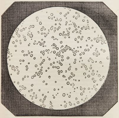
Fig. 40 – Thin Disk of Cow’s MilkArtist
Donné, Alfred (French, 1801-1878)Publication
The Museum of Science and ArtDate
1855 plate (1845 negative)Process
Engraving (from photograph)Image Size
9 x 9 cm
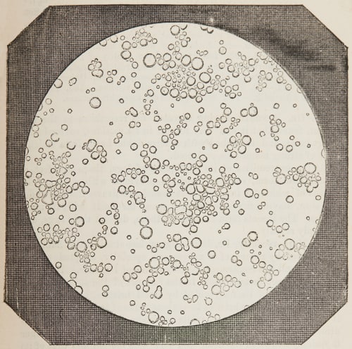
In 1844 and 1845 French physician Alfred François Donné published Cours de microscopie compémentaire des études médicales in Paris. The folio atlas of plates, which appeared one year after the text, included twenty plates showing engraved images of 86 micro-daguerreotypes taken by medical student, later physicist Léon Foucault. Because daguerreotypes were unique images they could not be duplicated by a photographic process like prints from photographic negatives, and had to be engraved for reproduction by printing.
Donné, a French public health physician, began teaching his pioneering course on medical microscopy in 1837, a time when the medical establishment remained largely unconvinced of the microscope’s usefulness as a diagnostic and investigative tool. When Daguerre announced to the Académie des Sciences his “daguerreotype” process for creating finely detailed photographic images in July 1839, Donné immediately embraced this new art, and within a few months had created not only the first documented photographic portrait in Europe, but also the earliest method of preparing etched plates from daguerreotypes. Donné resolved to incorporate photography into his microscopy course, and in February 1840 he presented to the Académie his first photographic pictures of natural objects as seen through the microscope. “It was Alfred Donné who foresaw the helpful role that projections of microscopic pictures could play during lectures on micrography” [1]
Over the next few years Donné continued to refine his photomicrography methods with the help of his assistant, Léon Foucault (who would go on to have a distinguished career as a physicist). Donne’s and Foucault’s work was the first biomedical textbook to be illustrated with images made from photomicrographs. Among its noteworthy images are the first microphotographs of human blood cells and platelets, and the first photographic illustration of Trichomonas vaginalis, the protozoon responsible for vaginal infections, which Donné had discovered in 1836.
In 1855 Donne and Foccult’s photo-micrograph engravings were reproduced in The Museum of Science and Art. From the accomanying text…
This fictitious application of the photographic art, to the promotion of natural science, after some experimental test, more or less successful, was first carried out, so as to be available for the practical purposes of science, by Dr. Donné assisted by M. Leon Foccult, in 1845. In the year Dr. Donné published an atlas to illustrate his course on microscopic anatomy and physiology, which had appeared in the previous year, consisting of twenty plates, on each of which were four microscopic engravings made from daguerreotype plates which had been produced in the manner above described. I avail myself gladly of the kind permission of the authors of this work, and of Mr. Bailliére, its publisher, to reproduce four of these engravings upon the scale on which they are given by the authors." P. 99
References
[1] Dreyfus, Some Milestones in the History of Hematology, pp. 38-40, 54-56, 76-78
Norman, Jeffery, History of Information https://www.historyofinformation.com/image.php?id=3394 cited 1/22/23
Frizot, A New History of Photography, p. 275.
Gernsheim & Gernsheim, The History of Photography 1685-1914, pp. 116, 539.
Hannavy, Encyclopedia of Nineteenth-Century Photography, Vol. 1, p. 1120.
Thorburn, “Alfred François Donné, 1801-1878, discoverer of Trichomonas vaginalis and of leukaemia,” British Journal of Venereal Disease 50 (1974) 377-380.